Anatomy Of Rib Cage Area - Rib Cage Muscles And Tendons / Anatomy Of The Sinew ... - The body of the horse, enclosing the rib cage and the major internal organs
Anatomy Of Rib Cage Area - Rib Cage Muscles And Tendons / Anatomy Of The Sinew ... - The body of the horse, enclosing the rib cage and the major internal organs. It has a roughened area on its upper surface, from which the serratus anterior muscle originates. The rib cage is collectively made up of long, curved individual. Hepar (in ancient greek) = liver An inhalation is accomplished when the muscular diaphragm, at the floor of the thoracic cavity, contracts and flattens, while the contraction of intercostal muscles lift the rib cage up and out. See thoracic spine anatomy and upper back pain
The area where the saddle sits, beginning at the end of the withers, extending to the last thoracic vertebrae (colloquially includes the loin or coupling, though technically incorrect usage) barrel: Quizzes are the secret to your success! In the mammary line (an imaginary vertical line from the right breast nipple), the liver extends from the 5th rib to the bottom of the rib cage 9. Jul 08, 2021 · the best way to learn anatomy is to repeat as much as you can. The curve becomes most prominent at the costal angle, which is when the rib turns anterolaterally.
Medical poster Anatomy print Rib cage art Macabre print ... from i.pinimg.com It encloses the thoracic cavity, which contains the lungs. The body, or shaft, of the rib is thin, flat and curved. The rib cage is collectively made up of long, curved individual. Hepar (in ancient greek) = liver In the mammary line (an imaginary vertical line from the right breast nipple), the liver extends from the 5th rib to the bottom of the rib cage 9. Rib 2 is thinner and longer than rib 1, and has two articular facets on the head as normal. An inhalation is accomplished when the muscular diaphragm, at the floor of the thoracic cavity, contracts and flattens, while the contraction of intercostal muscles lift the rib cage up and out. It is formed by the 12 thoracic vertebrae, 12 pairs of ribs and associated costal cartilages and the sternum.
Mar 20, 2015 · the ribs partially enclose and protect the chest cavity, where many vital organs (including the heart and the lungs) are located.
Rib 2 is thinner and longer than rib 1, and has two articular facets on the head as normal. Jul 08, 2021 · the best way to learn anatomy is to repeat as much as you can. See thoracic spine anatomy and upper back pain The human rib cage is a component of the human respiratory system. It is formed by the 12 thoracic vertebrae, 12 pairs of ribs and associated costal cartilages and the sternum. The thoracic spine is basically a strong cage and it is designed to protect the vital organs of the heart and lungs. It encloses the thoracic cavity, which contains the lungs. Quizzes are the secret to your success! The curve becomes most prominent at the costal angle, which is when the rib turns anterolaterally. In the mammary line (an imaginary vertical line from the right breast nipple), the liver extends from the 5th rib to the bottom of the rib cage 9. Its attachment to the rib cage affords the thoracic region of the spinal column greater stability and strength. It has a roughened area on its upper surface, from which the serratus anterior muscle originates. The area where the saddle sits, beginning at the end of the withers, extending to the last thoracic vertebrae (colloquially includes the loin or coupling, though technically incorrect usage) barrel:
Quizzes are the secret to your success! It encloses the thoracic cavity, which contains the lungs. The body, or shaft, of the rib is thin, flat and curved. The costal angle also marks the attachment for some of the deep back muscles to. The thoracic cage takes the form of a domed bird cage with the horizontal bars formed by ribs and costal cartilages.
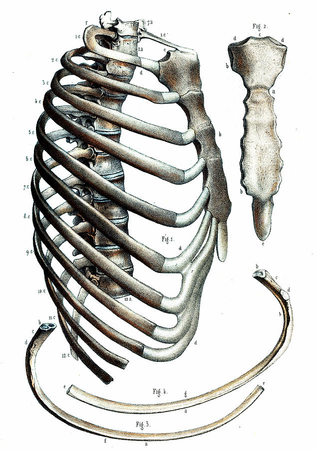
Human rib cage, side view | Рисунки, Тело from i.pinimg.com The thoracic spine is basically a strong cage and it is designed to protect the vital organs of the heart and lungs. See thoracic spine anatomy and upper back pain It has a roughened area on its upper surface, from which the serratus anterior muscle originates. The human rib cage is a component of the human respiratory system. It is supported by the vertical sternum or. The body, or shaft, of the rib is thin, flat and curved. The firm attachment of the rib cage at each level of the thoracic spine provides stability and structural support to the upper back and allows very little motion. The rib cage is collectively made up of long, curved individual.
Quizzes are the secret to your success!
Jul 08, 2021 · the best way to learn anatomy is to repeat as much as you can. The curve becomes most prominent at the costal angle, which is when the rib turns anterolaterally. The firm attachment of the rib cage at each level of the thoracic spine provides stability and structural support to the upper back and allows very little motion. The rib cage is collectively made up of long, curved individual. See thoracic spine anatomy and upper back pain The body, or shaft, of the rib is thin, flat and curved. It is formed by the 12 thoracic vertebrae, 12 pairs of ribs and associated costal cartilages and the sternum. It has a roughened area on its upper surface, from which the serratus anterior muscle originates. Jul 29, 2021 · the thoracic cage (rib cage) is the skeleton of the thoracic wall. The thoracic spine is basically a strong cage and it is designed to protect the vital organs of the heart and lungs. It encloses the thoracic cavity, which contains the lungs. Hepar (in ancient greek) = liver The costal pleura keeps it separated from the ribs and the deepest intercostal muscles (muscles running between the ribs, keeping the rib cage flexible) 7.
The costal angle also marks the attachment for some of the deep back muscles to. Jul 29, 2021 · the thoracic cage (rib cage) is the skeleton of the thoracic wall. See thoracic spine anatomy and upper back pain The costal pleura keeps it separated from the ribs and the deepest intercostal muscles (muscles running between the ribs, keeping the rib cage flexible) 7. The firm attachment of the rib cage at each level of the thoracic spine provides stability and structural support to the upper back and allows very little motion.
Anatomy Of Human Rib Cage - Anatomy - Human Rib Cage by ... from images.fineartamerica.com The curve becomes most prominent at the costal angle, which is when the rib turns anterolaterally. The costal angle also marks the attachment for some of the deep back muscles to. Mar 20, 2015 · the ribs partially enclose and protect the chest cavity, where many vital organs (including the heart and the lungs) are located. The firm attachment of the rib cage at each level of the thoracic spine provides stability and structural support to the upper back and allows very little motion. It is formed by the 12 thoracic vertebrae, 12 pairs of ribs and associated costal cartilages and the sternum. The thoracic spine is basically a strong cage and it is designed to protect the vital organs of the heart and lungs. See thoracic spine anatomy and upper back pain Jul 08, 2021 · the best way to learn anatomy is to repeat as much as you can.
Mar 20, 2015 · the ribs partially enclose and protect the chest cavity, where many vital organs (including the heart and the lungs) are located.
The human rib cage is a component of the human respiratory system. Jul 08, 2021 · the best way to learn anatomy is to repeat as much as you can. Sep 02, 2017 · it covers the largest area among the three surfaces of the lung 27. Hepar (in ancient greek) = liver The curve becomes most prominent at the costal angle, which is when the rib turns anterolaterally. The body of the horse, enclosing the rib cage and the major internal organs The surface between the left and right lungs, it houses the hilum. The costal angle also marks the attachment for some of the deep back muscles to. Rib 2 is thinner and longer than rib 1, and has two articular facets on the head as normal. The area where the saddle sits, beginning at the end of the withers, extending to the last thoracic vertebrae (colloquially includes the loin or coupling, though technically incorrect usage) barrel: The thoracic cage takes the form of a domed bird cage with the horizontal bars formed by ribs and costal cartilages. It has a roughened area on its upper surface, from which the serratus anterior muscle originates. The body, or shaft, of the rib is thin, flat and curved.
Hepar (in ancient greek) = liver anatomy of rib cage. Sep 02, 2017 · it covers the largest area among the three surfaces of the lung 27.


0 Komentar